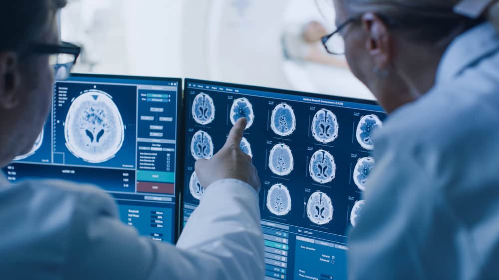
Artificial intelligence in imaging used to feel like patchwork: one small algorithm to flag lung nodules, another to clean noisy MRI slices, a third to suggest boilerplate text. Now, a supersized “foundation” model can handle all three jobs — often at once — because it has learned the shared grammar of pixels and prose from millions of images and their matching reports.
The jump from point solutions to all-purpose networks happened fast. Three forces pushed it forward: (1) public PACS archives that finally topped the petabyte mark; (2) transformer architectures that treat an image the way GPT treats a sentence; and (3) GPUs-on-demand that let research hospitals pre-train without buying a super-computer. The result is a model that can walk into any modality and still speak radiology.
Equally important, the shift has caught the eye of policymakers and payers. Germany’s 12-site PRAIM project demonstrated that an AI-backed reader can detect 17.6 percent more breast cancers without impacting recall rates, providing regulators with a concrete benchmark for efficacy. In the US, the FDA’s January 2025 draft guidance now frames foundation models as clinical decision-support, not merely study add-ons — a sign that broad deployment is no longer hypothetical.
A second set of eyes, only sharper
Radiologists have always double-checked one another, but the foundation model adds a tireless partner that never blinks. In the PRAIM rollout, software boosted the detection rate to 6.7 cancers per 1,000 screens, up from 5.7 in the human-only arm, while slightly lowering false alarms.
That same “extra set of eyes” helps with quality control. A multicenter study of 3,469 chest X-ray addenda found that an auditing algorithm identified 96% of mislabeled or missed findings, flagging errors long after the attending physician had signed out.
The broader payoff is consistency. By pooling patterns from hundreds of hospitals, the model narrows the performance gap between new residents and senior thoracic specialists so that everyone starts from the same statistical baseline, and then layers judgment on top. That democratization matters most at night and in smaller facilities, where a single reader may cover multiple modalities on their own.
See also: Why the Radiology Community is Eager to Embrace AI
Faster results when every minute counts
Speed gains can decide outcomes in trauma bays and stroke suites. A Mass General Brigham trial showed that AI-drafted chest X-ray reports cut the median reading time from 34 seconds to 19 seconds — a 42 percent workflow boost — while raising pleural-lesion sensitivity nearly ten points. Similarly, on busy mammography days, the PRAIM workflow allows radiologists to spend 43 percent less time on routine exams, reallocating those hours to suspicious cases and real patient conversations.
AI triage for large-vessel-occlusion stroke isn’t theoretical either. According to multiple ISC 2025 and UC Davis reports, hospitals that deployed Viz.ai’s alert system saw a 30-60 minute reduction in door-in-door-out and door-to-needle metrics, translating to meaningful gains in neurological function and a full year less disability for the average patient in real-world cohorts worldwide.
Personalized imaging for personalized care
Precision medicine begins with precise maps. Interactive contour tools, built on the Segment Anything model, let oncologists rough in a glioma border and watch the network refine it to sub-millimeter accuracy. A 2024 study reported a 3-D Dice score of 0.87 for low-grade tumors and 0.92 after multi-plane fusion, good enough for first-pass radiotherapy planning.
Because the backbone is modality-agnostic, fine-tuning it for liver, prostate, or cardiac volumes takes days, not months. Surgeons walk into the operating room with clearer virtual boundaries, and interventionalists steer catheters with real-time margin updates.
The same network now optimizes radiation dose by predicting contrast kinetics and organ motion, programming scanner parameters on the fly, and shaving 15–25 percent off cumulative exposure in pediatric protocols without sacrificing signal-to-noise.
Spotting disease before symptoms even appear
Early detection may be the biggest prize. In PRAIM, AI identified micro-calcifications that humans often flagged months later, adding one confirmed cancer per 1,000 screens. Vision-language models trained on longitudinal CT pairs already identify millimetric lung nodules that grow just enough to matter, giving pulmonologists more time to biopsy or monitor them.
Beyond big iron scanners, handheld ultrasound probes with embedded AI now beat expert readers by nine percentage points in spotting pulmonary tuberculosis in low-resource clinics — proof that sub-clinical signs aren’t limited to radiology suites.
Always learning, always transparent
Foundation models are never static. Each case teaches the next one, and that adaptability comes with obligations. The January 2025 FDA draft requires lifecycle monitoring, bias audits, and text-based rationales, pushing vendors to expose saliency maps and uncertainty scores with every release.
Hospitals, for their part, now demand change logs and algorithmovigilance dashboards before signing purchase orders, integrating software surveillance into quality-assurance rounds.
Privacy-preserving federated learning makes that evolution possible without shipping protected data off-site. A 2024 multicenter brain tumor project trained a segmentation model across nine institutions and still achieved a Dice score of 0.899 — within one point of the centralized benchmark — without patient data ever leaving local firewalls.
The road ahead
RSNA reviewers predict that next-gen networks will ingest EHR notes, genomics, and wearable feeds, turning radiology from a snapshot to a running commentary on a patient’s health story. And edge hardware is following suit.
Device makers already embed triage algorithms into portable MRI and CT units, allowing rural clinicians to prioritize high-risk scans before sending them to the cloud. In maternal care, Philips’ AI-guided handheld ultrasound — now backed by a major 2024 grant — aims to reduce global obstetric mortality by delivering anomaly screening to villages that have never had local access to a radiologist.
Foundation models won’t replace radiologists, but they are already redrawing the job description. By catching faint signals, shaving minutes off critical paths, tailoring maps to unique anatomies, and flagging trouble long before symptoms appear, these neural generalists let physicians spend more time doing what only humans can: weighing trade-offs, reassuring families, and deciding what happens next, face-to-face.
The best future may look less like “man versus machine” and more like an orchestra: the model handles the steady rhythm of detection and measurement, while clinicians work independently on the most challenging passages. Together, the model and clinicians will produce a sound that neither could have managed alone.

 Best Practices for Deploying and Scaling Industrial AIArtificial Intelligence (AI) is transforming industrial operations, helping organizations optimize workflows, reduce downtime, and enhance productivity. Different industry verticals leverage AI in unique ways.Link to The Center for Adaptive Edge Intelligence
 The Center for Adaptive Edge IntelligenceAdaptive edge intelligence brings real-time decision-making to the point of data creation, whether from sensors, machines, or cameras.Link to The Value of Vehicle Electrification
 The Value of Vehicle ElectrificationElectric vehicles (EVs) present automakers with many design, engineering, and manufactu ring challenges.Link to Accelerating Manufacturing Digital Transformation with Industrial Connectivity and IoT
 Accelerating Manufacturing Digital Transformation with Industrial Connectivity and IoTDigital transformation is empowering industrial organizations to deliver sustainable innovation, disruption-proof products and services, and continuous operational improvement.Link to Smart Manufacturing for Automotive
 Smart Manufacturing for AutomotiveLeading a transportation revolution in autonomous, electric, shared mobility and connectivity with the next generation of design and development tools.Link to Center for Data Pipeline Automation
 Center for Data Pipeline AutomationAs businesses become data-driven and rely more heavily on analytics to operate, getting high-quality, trusted data to the right data user at the right time is essential.Link to Center for Automated Integration
 Center for Automated IntegrationThe goal of automated integration is to enable applications and systems that were built separately to easily share data and work together, resulting in new capabilities and efficiencies that cut costs, uncover insights, and much more.Link to Continuous Intelligence: Insights
 Continuous Intelligence: InsightsDigital transformation requires continuous intelligence (CI). Today’s digital businesses are leveraging this new category of software which includes real-time analytics and insights from a single, cloud-native platform across multiple use cases to speed decision-making, and drive world-class customer experiences.