


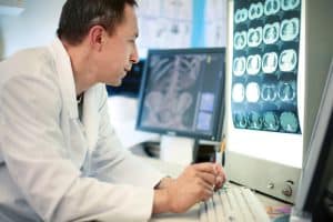


The radiology community is eager to embrace artificial intelligence since we believe that it has the potential to vastly improve patient outcomes.
AI will create a “paradigm shift,” similar to the introduction of MRI nearly forty years ago and picture archiving and communication systems (PACS) twenty years ago.
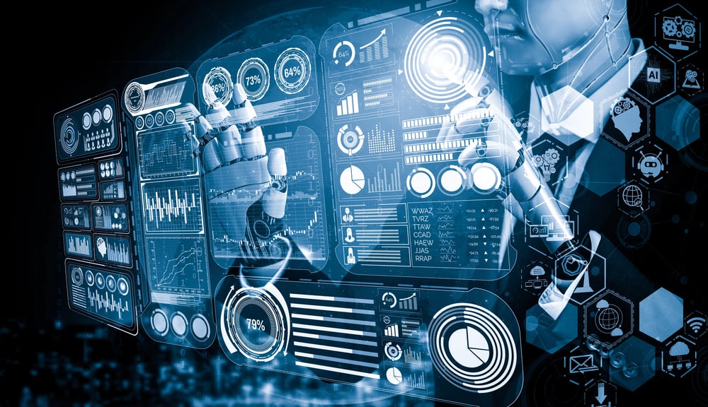
Artificial intelligence has begun to impact the practice of diagnostic radiology in profound ways including the ability to evaluate brain tumors, strokes and multiple sclerosis on MRI studies, diagnose pancreatic cancer and intracranial hemorrhage on CT studies, calculate bone age on hand radiographs, self-correct diagnostic reports, and triage worklists based on pre-selected criteria. Advanced AI software can compare the findings on a current medical image with those from prior imaging studies to look for responses to therapy, new lesions or acute changes.
See also: What’s the potential for real-time analytics in healthcare?
AI software more accurately measures the size and volume of neoplasms and demyelinating lesions than is possible by human measurements and calculations. Such software relieves the radiologist of repetitive tedious tasks and serves as a useful risk-management tool. Medical oncologists have long wished to know the true volume of tumors for the purposes of fine-tuning chemotherapy– thanks to AI software this is now readily available.
When comparing current and prior medical imaging studies, radiologists can now analyze co-registered images which show cross-sectional slices in the same plane, scale, thickness and angulation. Such co-registered images help avoid interpretation errors from changes that can occur when the studies differ by manufacturer, magnetic field strength and/or resolution.
The Kuiper belt object Pluto was difficult to locate until astronomers used a device called a “blink comparator” to rapidly alternate celestial photographs obtained months apart to look for differences in the night sky.
Similarly, medical images can now be rapidly alternated using digital computer methods to cause pathological changes to “flicker” and be more reliably seen and counted. AI software makes specialized maps of these changes, using colors and color intensity to indicate the type and degree of alterations of lesions.
Pharmaceutical companies are beginning to use medical imaging diagnostic AI software to measure therapeutic efficacies. This has the potential to improve the reliability and to reduce the time to conduct clinical trials, potentially bringing drugs to market sooner and reducing the overall cost of pharmaceutical development. If a new drug is found by AI techniques to be ineffective earlier in a clinical trial than was possible previously, fewer patients may be negatively impacted.
The field of radiology is one often cited as likely to be “disrupted” in the near future by artificial intelligence. We actually see this as a great opportunity for our specialty and we are historically used to accommodating positively with disruptions of similar magnitude. Radiologists are faced with ever-increasing workload burdens, tighter reporting deadlines and new, outcome-based payment models which shift reimbursement risks from the insurer to the interpreter. Those of us who lack AI tools will soon be at a great disadvantage.
At the time PACS systems were introduced we could for the first-time display images on electronic monitors rather than photographic film and there were tremendous efficiencies generated.
Radiographs and cross-sectional studies along with their comparison images could now be chosen from an electronic worklist rather than hunting in file rooms, lockers, or automobile trunks for misplaced x-rays. CT and MRI studies containing hundreds of images could now be displayed rapidly and in useful, novel formats. This resulted in an approximately 30%-time reduction. The introduction of artificial intelligence software to medical imaging theoretically could have similar performance gains and the potential for workforce reduction.
However, as we saw when PACS was introduced, there has been a continuous and unabated increase in the demand for medical imaging services and the number of studies keeps increasing.
Radiology, like all of medicine, is incredibly complex and nuanced. Radiologists will assume increasingly more “cognitive” and “patient-focused” roles as AI software relieves us of mundane activities that it can perform more quickly and accurately.
A radiographic differential diagnosis, unlike numerical findings such as blood pressure measurements or serum glucose levels, has deep significance on many levels, all of which require careful communication, follow-up and therapy recommendations, which increasingly are based on genomics and tailored individualized needs. The human element that a radiologist can provide will be more essential than ever.